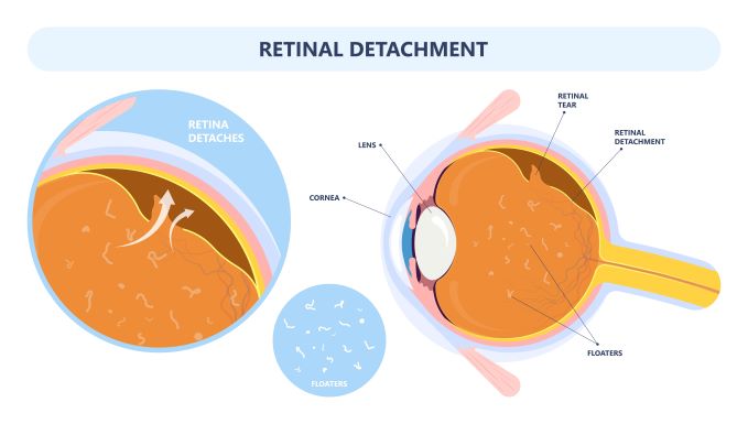
The retina plays a vital role in your vision and is located on the back wall of the eye. The retina’s primary function is to convert light into the images you see around you. As long as the retina is attached, it should work correctly. However, if the retina becomes detached from the back wall of the eye, this would require immediate attention from a retinal specialist. A detached retina is seemingly painless, so how do you know if you are experiencing a retinal detachment?

Flashes and Floaters
Flashes in your vision might appear like lightning bolts or stars. Floaters appear like opaque specks, waves, or cobweb-like lines in your visual field. The gradual onset of flashes and floaters is relatively common, especially for older patients or patients diagnosed with diabetes. However, an increased amount of these visual phenomena could signify a detached retina requiring immediate medical attention.
Decreased Vision
Another tell-tale symptom of a retinal detachment is the appearance of a dark shadow or curtain in your visual field. This dark shadow or gray curtain can appear in your peripheral vision, or at the top or bottom of your visual field. Once it appears, it will usually grow larger over time. This is a sign that the retina has already begun to detach from the back of the eye, and should be attended to immediately.
Risk Factors
Aging is the most common risk factor in experiencing a retinal detachment, but there are a few other risk factors as well:
- Family history of retinal detachments
- Myopia (nearsightedness)
- Previous eye surgery
- Previous retinal detachment in the other eye
- Trauma (blunt-force injury to the eye)
Diagnosis and Treatment
The ophthalmologist will dilate your eyes to widen your pupils in order to diagnose a retinal detachment. From there, a special lens is used to closely examine the retina. If your symptoms result from a retinal tear rather than a retinal detachment, then laser therapy or cryopexy is performed to seal the tear. However, more advanced tears and retinal detachments require surgery. Surgery is typically performed within a few days of the appointment.
There are a few surgical options depending on the extent of your detachment and which treatment is best for you. These include:
- Scleral buckle – a silicone band holds the retina in place, and a laser is used to seal the tear
- Vitrectomy – the vitreous is removed, and a gas bubble is inserted to push the retina back in place
- Pneumatic retinopexy – a gas bubble is injected into the vitreous, pushing the retina back in place
In both a vitrectomy and pneumatic retinopexy, the gas bubble and any fluid collected underneath the retina during its detachment gets reabsorbed into the body. After surgery, most people have vision close to what they had before the tear or detachment.
Retinal detachments are serious. If left unchecked and untreated, permanent vision loss can occur. If you experience any symptoms related to a tear or detachment, immediately call our office at 954-741-5555 to schedule an appointment with one of our experienced retinal surgeons: Dr. Burgess, Dr. Lara, or Dr. Villate.


