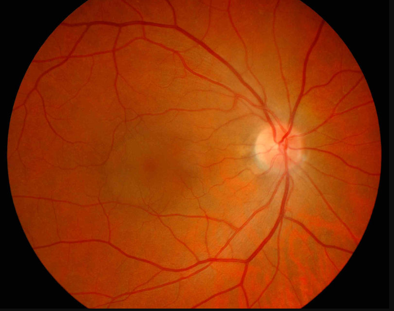
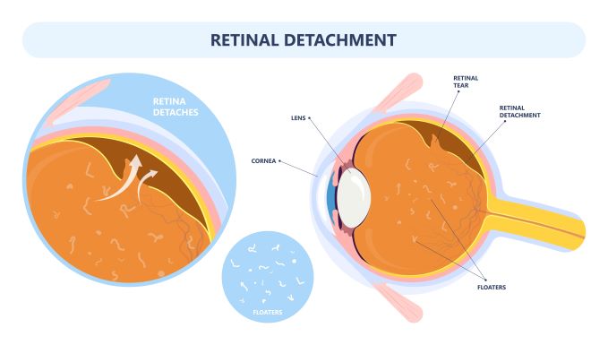
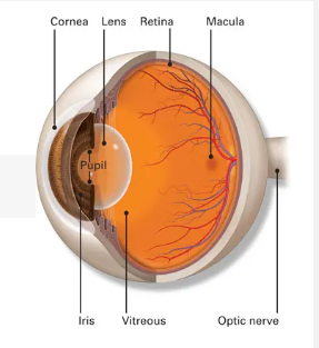
Central Serous Retinopathy (CSR) sometimes referred to as central serous choroidopathy, is an eye disease that causes visual impairment and usually occurs in one eye and is often temporary. Characterized by leakage of fluid under the retina, CSR has the propensity to accumulate under the central macula when active. Fluid leakage is believed to come from the choroid (a tissue layer with blood vessels under the retina). When tiny areas of the retinal pigment epithelium (RPE) become defective, fluid builds up and accumulates under the RPE – this causes a small detachment to form under the retina. CRE usually affects just one eye, but has the possibility of affecting both eyes.
Symptoms of CSR
- Distorted, blurred, or dim vision
- Blind spot in central vision
- Distortion of straight lines in the affected eye
- White objects may appear to have a brownish tinge or may be duller in color
- Objects may appear to be smaller or further away than they actually are
Interestingly, men are more likely to develop central serous choroidopathy than women – especially in their 30s to 50s. And stress is a major risk factor. There are even studies that suggest that people with “type A” aggressive personalities that are under a lot of stress may develop central serious retinopathy more often than those with less stressful lives. Other risk factors include high blood pressure (hypertension), the consumption of caffeine, use of steroids, and working-aged patients with occupations that demand high levels of visual acuity.
CSR Diagnosis
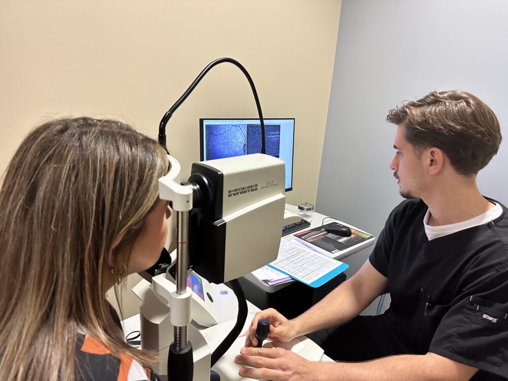
In order to diagnose whether or not you have central serous retinopathy, your ophthalmologist will dilate your eyes to examine your retina. If CSR is suspected, your ophthalmologist will then take photographs of your eye using fluorescein angiography and optical coherence tomography (OCT).
During fluorescein angiography, a fluorescent dye is injected into the patients arm so that it reaches the bloodstream and then highlights the blood vessels in the back of the eye so they can be photographed. Your doctor is then able to view any abnormal or leaky blood vessels to determine whether you have CSR. Fluorescein angiography also helps to determine the extent and progression of CSR and helps target specific treatment areas.
Indocyanine green (ICG) angiography may also be used for detection of central serous retinopathy. The difference between ICG and FA is that the dyes are different. Your ophthalmologist may choose to use either of these imaging methods, depending on your unique condition.
OCT scanning may also be used by your ophthalmologist to obtain a cross-section picture of your retina to measure retinal thickness and detect swelling of the retina.
CSR Treatment Options:
- Focal laser photocoagulation – pinpoints areas of leaking on fluorescein angiography
- Photodynamic therapy – targets the choroidal circulation
- Anti-VEGF – used for upregulation of tight junctions between endothelial cells and reduction of vascular fenestrations
Most central serous retinopathy cases resolve spontaneously within 3 to 4 months. Half of the patients who have had CSR will have a reoccurrence. Regular follow-up examinations are important to prevent long-term fluid accumulation that can lead to permanent vision loss.
