Retinal imaging technology has revolutionized the diagnosis, management, and treatment of various ocular conditions. This new technology offers a non-invasive and comprehensive view of the retina. This enables practitioners to detect, monitor, and treat a myriad of ocular diseases and disorders with precision. Many benefits in enhancing ocular healthcare have been made through advanced retinal imaging.
Understanding Retinal Imaging:
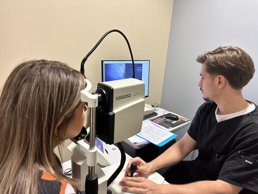
Retinal imaging encompasses a spectrum of imaging modes aimed at capturing high-resolution images of the retina. This is the delicate neural tissue lining the back of the eye. These imaging techniques utilize advanced optics, digital cameras, and sophisticated software algorithms to visualize the intricate structures of the retina. This includes the optic nerve head, macula, retinal vasculature, and peripheral retina. Common modalities of retinal imaging include: fundus photography, optical coherence tomography (OCT), fluorescein angiography, and fundus autofluorescence imaging.
Five Benefits of Retinal Imaging:
- Early Detection of Ocular Pathologies:
Retinal imaging facilitates the early detection of various ocular pathologies. These include diabetic retinopathy, age-related macular degeneration (AMD), glaucoma, retinal vascular diseases, and retinal detachment. By capturing detailed images of the retina, clinicians can identify subtle changes indicative of disease onset or progression. Which enables timely intervention and preventive measures to preserve vision.
- Objective Disease Monitoring:
Retinal imaging provides objective and quantitative data for monitoring disease progression and treatment response. The optical coherence tomography (OCT), enables precise measurement of retinal thickness, macular volume, and other morphological parameters. This aids in the evaluation of therapeutic effectiveness and disease management strategies.
- Personalized Treatment Planning:
With retinal imaging, clinicians can tailor treatment plans to the individual characteristics of each patient’s retina. By analyzing high-resolution images and identifying specific anatomical features such as: macular edema, subretinal fluid, or drusen deposits. Practitioners can devise personalized treatment regimens including: intravitreal injections, laser therapy, or surgical interventions.
- Enhanced Patient Education:
Retinal imaging facilitates patient education and engagement by visually illustrating ocular conditions and treatment options. Clinicians can utilize retinal images to explain disease processes, highlight treatment goals, and demonstrate the rationale behind recommended interventions. This helps to empower patients to actively participate in their ocular health management.
- Advancements in Research and Innovation:
Retinal imaging serves as a cornerstone for research and innovation in the field of ocular health. By facilitating non-invasive visualization of retinal anatomy and pathology, this technology fuels the development of diagnostic tools, therapeutic interventions, and prognostic markers, driving advancements in the understanding and management of ocular diseases.
Retinal imaging represents a paradigm shift in the diagnosis, management, and treatment of ocular diseases, offering insight into the structure and function of the retina. With its benefits for early disease detection, personalized treatment planning, and patient education. As technology continues to evolve, further innovations in retinal imaging will continue to emerge in the future for more advanced testing in ocular healthcare.


