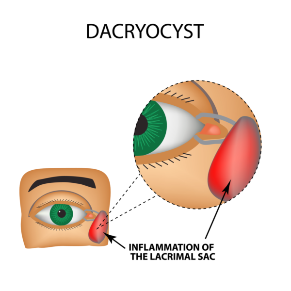
When the tear drainage system is either fully or partially blocked, it is a blocked tear duct. Tears that are unable to drain normally may result in wet, itchy, or infected eyes. People of all ages may experience symptoms and can possibly develop this condition.
Basic tears are produced in the conjunctiva lining the lids. Your lacrimal glands, positioned above each eye, produce most of your tears. Tears lubricate and protect the surface of your eye before draining through tiny ducts in the corners of your upper and lower eyelids.
Clogs can occur in any part of the tear drainage system. When this happens, your tears don’t drain properly, resulting in watery eyes and an increased risk of infection and inflammation in your eyes.
This condition can also be caused by congenital maldevelopment of the tear ducts. Oftentimes, acquired obstruction of the tear duct system could be caused by trauma, infection, nasal disease, and tumors in rarer cases.
When the tear ducts become blocked, bacteria could be trapped in the nasolacrimal sac, causing an infection named dacryocystitis.
Symptoms of this infection include:
- Inflammation, pain, and redness around the eye and nose.
- Recurrent infections of the eyes
- Mucus discharge from the eyes
- A mass in the inner corner of the eye
- Hazy vision (rare cases)
- Tears stained with blood
- Fever (rare cases)
Diagnosis of a tear duct entails a complete examination to rule out potential causes. The duct may be blocked at different parts of the system, requiring specific treatment. It’s also possible to get an X-ray or CT scan to confirm the obstruction and its location.
Sometimes, more than one treatment or procedure is needed before a blocked tear duct is fully opened. Antibiotics will almost certainly be prescribed if an infection is detected.
The surgical operation dacryocystorhinostomy is performed to treat most cases of clogged tear ducts in adults but may be found in children. By forming a new link between your lacrimal sac and your nose, this method allows tears to drain normally through your nose once more. This new path avoids the duct that empties into your nose (nasolacrimal duct), which is frequently clogged. In most cases, stents or intubation are placed in the new channel while it heals and then withdrawn three to six weeks later. The steps in this technique will differ depending on the severity of your tear duct obstruction.
Depending on the type of obstruction, a surgeon may consider establishing an entirely new channel from the inside corner of your eyes to your nose, bypassing the tear drainage system altogether. This reconstruction of your entire tear drainage system is called conjunctivodacryocystorhinostomy, a procedure in which a glass tube is used to bypass the obstruction.
In the rare case that a tumor is causing your blocked tear duct, biopsy and treatment of the growth may be necessary.
If you have experienced any symptoms associated with blocked tear duct disease, contact us to schedule an appointment.
Our very own Dr. Gil Epstein is a fellowship-trained, board-certified ophthalmic surgeon who performs all of the cosmetic and reconstructive oculoplastic procedures at the Fort Lauderdale Eye Institute. He is certified by the American Board of Ophthalmology and is a member of the American Society of Ophthalmic Plastic and Reconstructive Surgery.


