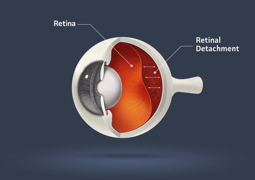
The retina is a layer of light-sensitive tissue that lines the inside of the eye and transmits visual signals to the brain via the optic nerve. A retinal detachment ensues when the retina is lifted or pulled from its natural position.

There are three types of retinal detachments:
Rhegmatogenous– The most common type of retinal detachment. A break or tear in the retina allows fluid to get under the retina and then separate it from the retinal pigment epithelium.
Tractional– Scar tissue on the retina’s surface causes the retina to lift up due to contraction.
Exudative– Fluid leaks into the area beneath the retina, but there are no tears or breaks in the retina. This detachment is typically caused by retinal diseases, including inflammatory disorders and trauma. A detached retina needs immediate surgical correction. Surgical repair procedures depend primarily on the patient’s diagnosis, the extent of damage, the cause of detachment, and the patient’s condition.
Pneumatic Retinopexy– Typically an office-based procedure that involves injecting an expanding gas bubble into the vitreous space inside the eye. Your doctor may use a freezing method known as cryotherapy in conjunction with a laser to seal the retina if additional treatment is needed.
Scleral buckling is typically carried out in an operating room. During this procedure, the retina is pulled back into place by a flexible silicone band that is wrapped around the eye. Depending on your health, it could be necessary to drain the fluid under the detached retina so that it can return to its natural position. This surgery’s fundamental advantage is avoiding additional retinal tugging, which enables the tear to heal gradually and prevents fluid from entering the sub retinal area.
A vitrectomy is carried out soon after laser surgery to remove the vitreous gel that is tugging on the retina. Three tiny incisions are made inside the eye during this technique to gain access to the inside and directly fix the separation. When a gas bubble is used, your body’s own fluid will gradually replace it. There will be a surgical procedure necessary to eliminate an oil bubble.
Depending on the procedure you’ve had to repair your retinal detachment, recovery normally takes between two and four weeks. Your doctor may put drops in your eye to prevent infection by preventing the pupil from opening widely or closing. You may also be requested to wear an eye patch for a few days.
You should stay away from flying or traveling at high altitudes if a gas bubble is put into your eye. Patients should refrain from strenuous activities during the first week of recovery, including watching television or engaging in other activities requiring prolonged screen viewing. Driving should be postponed until your vision returns to normal or until your ophthalmologist provides the all-clear. After a couple of weeks, you should be able to go back to work; however, rehabilitation may take longer.


