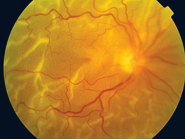
The retina is the light-sensitive layer of cells (tissue) that lines the inside of the eye and sends visual messages through the optic nerve to the brain. The optic nerve carries visual images from the back of your eye to your brain. A retinal detachment occurs when the retina is lifted or pulled from its normal position (the inner wall of the eye).
There are three types of retinal detachments:
- Rhegmatogenous – The most common type of retinal detachment. A break or tear in the retina allows fluid to get under the retina and then separates it from the RPE (retinal pigment epithelium).
- Tractional – Scar tissue on the retina’s surface causes the retina to separate due to contraction.
- Exudative – Fluid leaks into the area beneath the retina, but there are no tears or breaks in the retina. This type of detachment is typically caused by retinal diseases, including inflammatory disorders, injury to the eye and trauma.
A detached retina needs immediate surgical correction. There are several surgical procedures for retina repair, depending on the patient’s diagnosis, extent of damage, cause of detachment and patient condition.
Retinal Detachment Surgery Procedures
Pneumatic Retinopexy
Pneumatic retinopexy is typically and office-based procedure. It involves injecting an expanding gas bubble into the vitreous space inside the eye. The gas bubble gently pushes the retina into place against the back wall of the eye. If additional treatment is needed, your doctor may use a freezing device known as cryotherapy and laser to seal the retina.
Scleral Buckling
This procedure is typically performed in an operating room. With a scleral buckle, a flexible band composed of silicone is placed around the eye to counteract the force pulling the retina out of place. Depending on your condition, fluid under the detached retina may need to be drained to allow the retina to settle back into its normal position. The benefit of this procedure is to stop further pulling on the retina and cover the tear in the retina to prevent fluid from entering the subretinal space.
Victrectomy
Commonly performed in an operating room, a vitrectomy is performed to remove the vitreous gel which is pulling on the retina, and is sometimes performed following laser surgery. This procedure is done by making three micro incisions in the eye that allow access to the inside of the eye and repair the detachment directly. An oil or gas bubble may be used to keep the retina in place. When a gas bubble is used, your body’s own fluids will gradually replace it. And oil bubble will need to be removed with an additional surgical procedure at a later date.
Retinal Detachment Surgery Recovery
Depending on the type of procedure you’ve had to repair your retinal detachment, recovery typically takes anywhere from two to four weeks or more. Your doctor may put drops in your eye to keep the pupil from opening wide or closing and to prevent infection. You may also need to wear a patch over the eye for a few days.
When a gas bubble is used, it is recommended that you avoid flying or travelling at high altitudes. With other retinal detachment surgical procedures it is recommended during the first week of recovery that patients avoid strenuous activities, watching television, and any activity that requires staring at computer or cell phone screens for extended periods of time. Driving should be put on hold until your vision is back to normal or until your eye doctor gives you the green-light to go ahead and drive. Typically, you will be able to return to work after a couple of weeks. However, the recovery process is different for each patient.
photo credit: Community Eye Health


